

Superficial White Matter Imaging Biomarkers in Non-lesional Temporal Lobe Epilepsy: a multimodal study on microstructure pathology and clinically associated phenotypes
Temporal lobe epilepsy (TLE) is the most common focal epilepsy in adults. In about 30% of patients, MRI shows no identifiable lesion, even with dedicated epilepsy protocols. These non-lesional (MRI-negative) forms do not share the typical atrophy pattern of TLE with hippocampal sclerosis. Their underlying pathology, cognitive profile, and long-term course remain poorly understood, and imaging biomarkers able to capture them are still lacking.
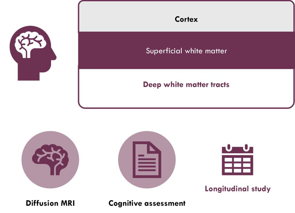
WINE focuses on the superficial white matter, the thin layer of short-range U-fibres lying just beneath the cortex. Standard MRI cannot see changes in this layer, but advanced post-processing can detect them. In a prospective longitudinal study, we compare patients with MRI-negative TLE, patients with hippocampal sclerosis, and healthy controls. We combine multimodal MRI (cortical morphometry, superficial and deep white matter microstructure, structural connectome, amygdala subnuclei) with a comprehensive neuropsychological assessment, repeated after 12–18 months.
Aim: test whether MRI-negative TLE comprises distinct phenotypes, identify early markers of cognitive decline, and support clinicians in a more personalised diagnostic and prognostic work-up.
- Funded by the Italian Ministry of Health, Ricerca Finalizzata 2024 – Starting Grant
- Duration: 2027–2030
- Principal Investigator: Alice Ballerini
- Supervisor: Prof. Anna Elisabetta Vaudano
- Host institution: Azienda Ospedaliero-Universitaria di Modena, in collaboration with the University of Modena and Reggio Emilia
Related publications:
- https://pubmed.ncbi.nlm.nih.gov/41873635/
- https://pubmed.ncbi.nlm.nih.gov/37531834/
- https://pubmed.ncbi.nlm.nih.gov/36213310/